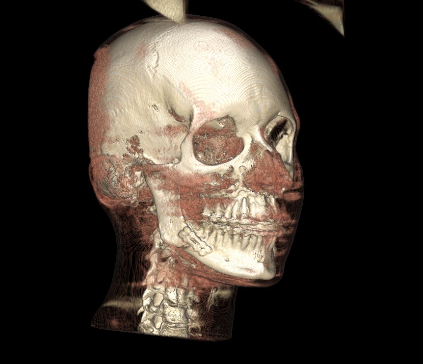
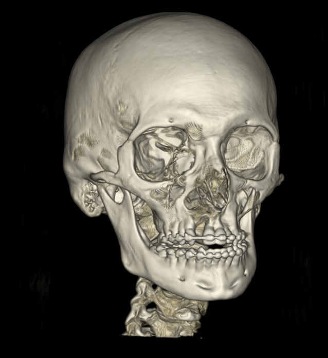
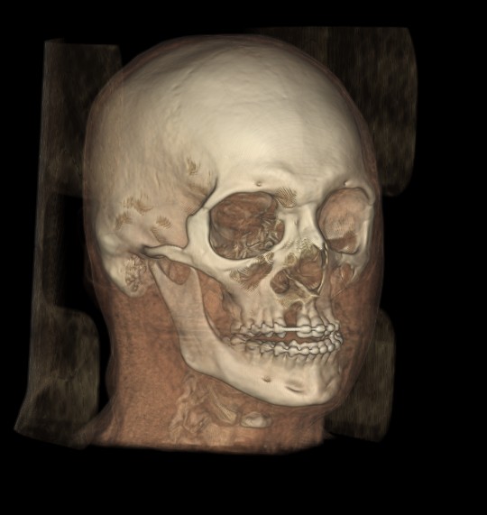
01 · CBCT
Cone beam computed tomography
CBCT is the anatomical backbone of every surgical plan and the reference frame for aligner and miniscrew cases. We require the raw DICOM dataset — not screenshots or exported renderings.
Technical
- Format: DICOM (single series, uncompressed)
- Voxel size: ≤ 0.4 mm for surgical, ≤ 0.5 mm for orthodontic
- Slice thickness: ≤ 0.4 mm
- FOV (surgical): full craniofacial, include cranial base, orbits, chin, cervical vertebrae
- FOV (orthodontic): dento-maxillary, include full dentition and TMJ bilaterally
Patient positioning
- Occlusion: centric relation, teeth in maximum intercuspation
- Head posture: natural head position, Frankfort plane horizontal
- Lips: relaxed, not retracted
- Tongue: in normal resting position
- Metal artefacts: minimise — remove jewellery, fixed prostheses where possible
02 · Intraoral scans
Digital impressions
Intraoral scans replace conventional impressions for all our setups. We accept exports from 3Shape TRIOS, iTero, Medit, Carestream and any system that can output clean STL/PLY.
Required files
- Upper arch — full arch, retromolar to retromolar
- Lower arch — full arch, retromolar to retromolar
- Bite registration — in centric occlusion
- Format: STL, PLY or OBJ (no proprietary containers)
Quality checklist
- No holes or missing surfaces on occlusal or incisal edges
- Clean gingival margins — retract tissue during scan
- Include 2–3 mm of gingiva for aligner cases
- Bite scan captures both arches in contact, not open
- Scans exported un-meshed / non-simplified if possible
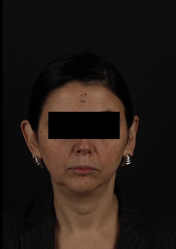
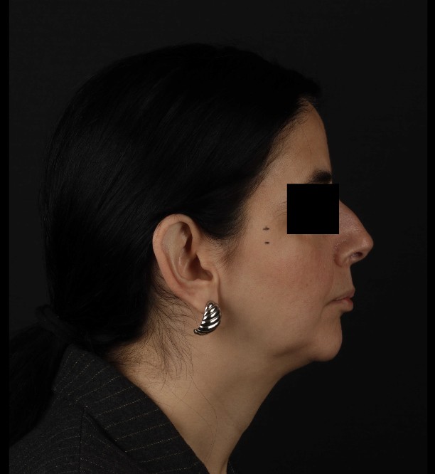
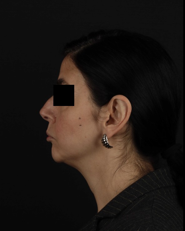
03 · Clinical photographs
Extraoral and intraoral photo set
Photographs capture soft tissue, aesthetics and expression — information CBCT and scans cannot provide. The standard set below is required for every case; surgical and smile design cases require additional views.
Extraoral (required)
- Frontal — relaxed lips
- Frontal — full smile
- Right profile — relaxed lips
- Right 3/4 — relaxed and smile
- Left profile & 3/4 for surgical cases
Intraoral (required)
- Frontal occlusion
- Right and left buccal segments in occlusion
- Upper and lower occlusal views with mirror
- Overjet view (profile, mouth slightly open)
Technical
- Format: JPG/PNG, minimum 2000 px long edge
- Background: neutral, non-distracting
- Lighting: even, no harsh shadows, flash or ring light
- White balance: calibrated; avoid warm/cool colour casts
- Orientation: Frankfort horizontal in profile views
Surgical extras
- Natural head position video (short clip)
- Repose and E-line reference
- Full body posture photo if relevant
- Dynamic smile video for smile design cases
04 · 2D radiographs
Panoramic and lateral cephalogram
Optional when a high-quality CBCT is available (we can derive these views), but helpful if already acquired as part of your standard records. Submit in original resolution.
- Panoramic radiograph — JPG/PNG, original resolution
- Lateral cephalogram — calibrated ruler or known measurement visible
- Hand-wrist film for growth assessment, when indicated
05 · Clinical prescription
The prescription makes the plan match your clinical intent.
We read every word. A clear prescription reduces revision rounds and yields a first-pass plan that already matches your objectives.
- Chief complaint and treatment goals
- Skeletal and occlusal priorities, any hard constraints
- Extractions / non-extraction decision, if already made
- Aligner, appliance, or surgical preferences
- Known anatomical or medical limitations
Download the current prescription templates from the downloads page.